Infectious diseases

Dr Linda Calabresi
Clinical Articles iconClinical Articles

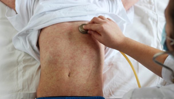
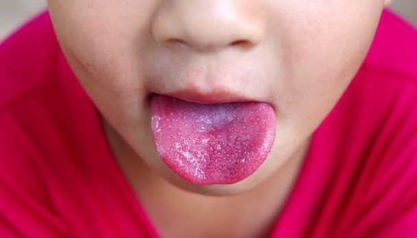
Scarlet fever is on the rise. According to the latest issue of The Lancet Infectious Diseases, cases of scarlet fever in the UK reached a 50 year high last year with a seven fold increase in new cases in the last five years. In addition, similar increases having been reported in a number of Asian countries including Vietnam, China, South Korea and Hong Kong. But public health authorities remain perplexed as to why the disease appears to be making a comeback. Detailed analysis of the causative organism shows different strains of the strep bacteria have been responsible for the UK and Asian outbreaks, so they are unsure if they are linked at all or whether the resurgence has to do with external factors such as the immune status of the population or environmental factors. So far it would seem that Australia is yet to be affected by this increased incidence however experts are warning we should not be complacent. Unlike in England, scarlet fever is not a notifiable disease in this country except in WA. And even in the UK, data suggests marked under-reporting. Scarlet fever is highly contagious and usually affects children under the age of 10, although it can occur in adults as well. While the bacterial infection, caused by Streptococcus pyogenes or group A Streptococcus (GAS) was a common cause of death in the 1800s, these days it is readily treated with antibiotics usually penicillin. However, failure to recognise the condition and treat it appropriately can lead to complications such as pneumonia, and liver and kidney damage. Children with the infection typically experience sore throat, headache and fever along with the characteristic popular pink-red rash that feels like sandpaper and the so-called strawberry tongue. Diagnosis is usually made via a throat swab. In an accompanying comment, Australian infectious diseases researchers Professor Mark Walker and Stephan Brouwer from the University of Queensland said, “Scarlet fever epidemics have yet to abate in the UK and northeast Asia. Thus, heightened global surveillance for the dissemination of scarlet fever is warranted.” In other words, be alert, people! Ref: Lancet Infect Dis 2017 Published Online November 27, 2017 http://dx.doi.org/10.1016/ S1473-3099(17)30693-X Online/Comment http://dx.doi.org/10.1016/ S1473-3099(17)30694-1

Dr Jenny Robson
Clinical Articles iconClinical Articles

Schistosomiasis, also known as bilharzia, is the second most prevalent tropical disease after malaria and is a leading cause of morbidity in many parts of the world. It is not uncommon in Australia because of the many travellers who visit endemic areas and swim or bathe in freshwater lakes and streams. Places commonly implicated include Lake Kariba and Lake Malawi in Africa. Immigrants and refugees from bilharzia endemic countries are also likely to present with untreated infection. With increasing travel to and migration from Africa and the Americas knowledge of the dangers and means of avoiding schistosomiasis is essential. Schistosomiasis is caused by trematodes of the genus Schistosoma. The principal schistosomes of medical importance, S japonicum, S mansoni, S mekongi (intestinal schistosomiasis) and S haematobium (urinary schistosomiasis), infect people who enter water in which infected snails (intermediate hosts) are living. The larval cercariae shed by the snail actively penetrate unbroken skin and develop into schistosomulae that migrate through the lungs to the liver where they mature into adults. Female worms lay eggs that pass through the vessels and tissues to the lumen of the gut or bladder (depending on localisation of worms). A proportion of eggs escape from the host and may be found in faeces or urine. The host's immune response to eggs that become lodged in the tissues is largely responsible for disease, Figure 1.  

Geographic distribution

This is governed by the distribution of the intermediate host snail. S haematobium                         Africa, Middle East, India (only Maharashtra) S japonicum                               Philippines, Indonesia (only Sulawesi), parts of China S mansoni                                   Africa, Middle East, some Caribbean Islands, parts of South America (Brazil, Surinam, Venezuela) S mekongi                                   Laos and Cambodia S intercalatum                           10 countries within the rainforest belt of West Africa.

At-risk groups

Owing to the absence of suitable snail hosts, transmission cannot occur in Australia. A history of overseas travel or residence is essential for this diagnosis. Chronic schistosomiasis is more likely to be seen in migrants and refugees from endemic areas. In Australia, where the definitive host is freshwater and marine birds, non-human trematodes may cause schistosomal dermatitis (cercarial dermatitis, swimmer's itch). Onset is usually within 15 minutes of skin contact with cercariae.

Clinical presentation

Disease due to schistosomiasis depends on the infecting species and the intensity of infection. Acute schistosomiasis occurs two to 12 weeks post infection and symptoms last for periods varying from one day to a month or more; recurrence of symptoms 2-3 weeks later is common. Between 40-95% of individuals, not previously exposed to infection, develop symptoms which include fever, malaise, headache, abdominal pain, diarrhoea and urticaria. Many have eosinophilia. After the initial acute onset, most become asymptomatic, although those with S haematobium infections may develop microscopic or macroscopic haematuria. Rare complications result from ectopic deposition of eggs in the spinal cord and brain. Most travellers are only mildly infected and are therefore often asymptomatic and unlikely to develop the severe manifestations of chronic schistosomiasis. Severe disease occurs in patients with heavy and prolonged infection. Hepatosplenomegaly, portal hypertension, ascites and oesophageal varices may result from intestinal schistosomiasis. And frank haematuria with varying degrees of impairment of the urinary bladder and ureters may occur with S haematobium infections.

Diagnosis

The prepatent period of S japonicum, S mansoni and S mekongi is 6-8 weeks, and for S. haematobium 10-12 weeks. Examination of faeces or urine before this time often yields false negative results. Similarly, with serology, testing too early may result in false negative results. Antibody development occurs slightly before eggs are detected. Eosinophilia (greater than 0.60 x10³/mL) is present in up to 80% of patients with infections; however, its absence does not exclude infection.

Parasitologic examination

Diagnosis is by demonstration of eggs of S japonicum, S mansoni and S mekongi  in faeces, or eggs of S haematobium in urine. At least two stool or urine specimens should be submitted for examination over a period of 10 days. Whilst eggs may be found in all specimens of urine, there is some evidence of a diurnal periodicity with a peak of excretion around midday. Collection of the terminal portion of urine collected between noon and 2 pm is therefore recommended. Schistosome eggs can also be demonstrated in rectal snips or bladder biopsies. Viability of eggs can be assessed if the biopsies are received fresh.

Serologic examination

At our laboratory, antibodies are detected by enzyme immunoassay (EIA) using purified egg S mansoni antigen. Antibodies to this antigen may be undetectable in the pre-patent period lasting 8-10 weeks. The test detects genus specific antibodies. In the absence of a diagnosis based on egg identification, travel history provides the best assessment of likely species.

Interpretation

Parasitologic Faeces is concentrated (modified formalin-ethyl acetate) and urine either centrifuged or filtered; all of the concentrate or sediment is examined. Because of the low sensitivity of these techniques, a negative faecal or urine examination does not exclude schistosomiasis. Microscopic examination of eggs enables the species of parasite to be determined. At least two examinations on different days are recommended. Serologic Schistosome serology cannot distinguish between past or current infection nor differentiate the species of infection. Clinical history and further investigations should be considered when establishing the diagnosis. Recent infections may be serologically negative.

Preventative measures

Cercariae can burrow through the mucosa of the mouth as well as through unbroken skin. All fresh water in endemic areas should be considered suspect, although snails tend to live in slow-flowing and stagnant waters, rather than in rapids and fast-flowing waters. If freshwater contact is unavoidable, bathing water should be heated to 50°C for five minutes or treated with iodine or chlorine as for the treatment of drinking water. Water can also be strained through paper filters, or allowed to stand for 2-3 days before use. This exceeds the usual life span of the cercariae. Of course, the container must be kept free of snails. High waterproof boots or hip waders are recommended if wading through streams or swamps. It is wise to carry a pair of rubber gloves to protect hands when contact with fresh water is anticipated. Vigorous towel drying, and rubbing alcohol on exposed skin immediately after contact with untreated water, may also help reduce cercarial penetration. Vegetables should be well cooked and salads avoided as these may have been washed in infected water, allowing cercariae to attach themselves to the leaves.

Treatment

Praziquantel (Biltricide) 20 mg/kg bodyweight every four hours for 2-3 doses depending upon the species is recommended. In travellers, this is likely to achieve cure rates in the order of 90%. Tablets are scored and available as a 600mg dose dispensed six per pack. In patients at risk of chronic disease, such as refugees and migrants, it is important to be aware of complications that may arise from chronic infection: liver fibrosis, portal hypertension and its sequelae, and colorectal malignancy in the intestinal forms; obstructive uropathy, superimposed bacterial infection, infertility and possibly bladder cancer.

Follow-up

Follow-up schistosomiasis serology is recommended in 12 to 36 months after treatment. Follow-up serology may differ between immigrants and returned travellers. Travellers may show a more rapid serological decline post-treatment due to a shorter duration of infection and lower parasite burden. Immigrants may even show a rise in titre within the first 6-12 months post-treatment. Persisting titres should not automatically justify retreatment, this should be based on symptoms, parasite identification or eosinophilia. Viable eggs may continue to be excreted for up to one month after successful treatment. Non-viable and degenerate eggs can be found in tissue biopsies for years after infection has occurred.
General Practice Pathology is a new regular column each authored by an Australian expert pathologist on a topic of particular relevance and interest to practising GPs. The authors provide this editorial, free of charge as part of an educational initiative developed and coordinated by Sonic Pathology.
Dr Linda Calabresi
Clinical Articles iconClinical Articles

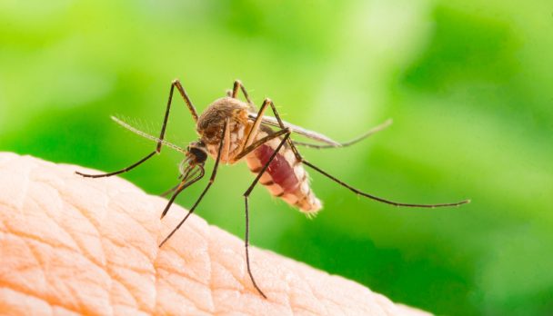
In what could represent a major blow to tourism in the region, the US Centers for Disease Control have, this week, issued a level 2 warning that mosquitoes in Fiji have been found to be infected with Zika virus and have transmitted the infection to humans. Because of the strong link between Zika virus infection and severe birth defects, the CDC is strongly advising against women who are pregnant or who are even planning on becoming pregnant travelling to the area. And as the virus can also be transmitted through the sex, the advice for pregnant women whose partner has travelled to Fiji is to use condoms or refrain from sex for the duration of the pregnancy. The warning also signals an alert for Australian doctors to consider Zika virus in patients who present with symptoms such as fever, rash and headache following travel to Fiji. However, one of the major problems in curtailing the spread of this virus has been the fact that infected adults may display very few if any symptoms and maybe unaware that have contracted the disease. What’s more an infected male can harbour the Zika virus in his semen for much longer than in other bodily fluids, so the CDC recommends that men travelling to a Zika-prone country, that now includes Fiji, avoid conceiving a child for six months after leaving the area or from the time they develop symptoms if they indeed do develop symptoms. Women clear the virus more quickly and therefore the recommendation from the CDC is that they avoid falling pregnant two months after potential exposure or from when symptoms appear, assuming their partner did not travel. For those people, including pregnant women who can’t avoid travel Fiji or other Zika-prone area, the CDC advises they take precautions to avoid mosquito bites and continue these precautions for three weeks after returning home. These include the use of specific insect repellents and the wearing of long-sleeved clothing. Ref: https://wwwnc.cdc.gov/travel/notices/alert/zika-virus-fiji

A/Prof Ian M. Mackay
Clinical Articles iconClinical Articles

This year, the number of laboratory-confirmed influenza (flu) virus infections began rising earlier than usual and hit historic highs in some Australian states. If you have been part of any gathering this winter, this is probably not news. States in the south-east (central and southern Queensland, New South Wales, Victoria, Tasmania and South Australia) are more inflamed by flu than those in the north and west. For example, Queensland has seen more hospital admissions than in the last five years, mostly among an older population, while younger demographics more often test positive without needing hospitalisation. Meanwhile, flu numbers in New Zealand and elsewhere in the Pacific have not matched the same elevated levels. But is Australia really experiencing the biggest flu season on record in 2017, or are we just testing more and using better tools? This is hard to answer for certain because the information we need is not usually reported until later and public databases only show the past five years. We can say for sure that 2017 is on track to be a historically big flu year.
Read more: Have you noticed Australia’s flu seasons seem to be getting worse? Here’s why

Really, a big flu season

Flu can be a nasty illness. Sometimes it’s deadly. Other times it can be mild. But even for cases that fall in the middle you may not be able to work for days, or you’ll have to look after ill children home from school, or visit the very sick who have been hospitalised. Years ago, detection of influenza viruses mostly relied on slow, finicky methods such as testing for virus in artificial cell cultures. But, in Australia today, most laboratories use either sensitive tools to detect viral gene sequences in samples from the patient’s airway, or less sensitive but rapid dipstick methods, where a special strip is placed in a sample to detect viral proteins. These tools have been in use since 2007 in the larger Australian laboratories, so it’s unlikely we are just seeing more positives in 2017. While newer versions of these tests are being rolled out this year, they are unlikely to detect more cases. Equally, it’s unlikely more people with suspected flu decided to change their behaviour in 2017 and get tested, compared to 2016, or the year before. As in all years, there are many people in the community with flu who don’t get tested. The proportion of people with flu who are tested likely remains roughly the same year to year. State-wide flu reports provide reliable, laboratory-confirmed results. By looking at them, we can also be confident that “man flu” and severe common colds aren’t contributing to this specific and large increase in flu. We’re very likely seeing a truly huge flu season.

Why so bad this year?

Flu, caused by infection with an influenza virus, is mostly a disease with an epidemic peak during July and August in non-tropical countries. Flu viruses are broadly grouped into two types: Influenza-A and Influenza-B. Influenza-B viruses have two main sub-types while the Influenza-A viruses are more variable. The Influenza-As you get each year are usually A/H3N2 (the main player so far this season) or A/H1N1, which lingers on from its 2009 “swine flu” pandemic. Multiple flu viruses circulate each year and serial infections with different strains in the same person in a single season are possible. H3N2 has played a big role in the past five flu seasons. When it clearly dominates we tend to have bigger flu seasons and see cases affecting the elderly more than the young. H3N2 is a more changeable beast than the other flu viruses. New variants can even emerge within a season, possibly replacing older variants as the season progresses. This may be happening this winter, driving the bigger-than-normal season, but we won’t know for certain until many more viruses are analysed. Outside winter, flu viruses still spread among us. This year, in particular, we’re being encouraged to get vaccinated even during the peak of flu season. Vaccines are a safe way to decrease the risk that we or loved ones will get a full-blown case of the flu. Yet Australian flu vaccination rates are low. Data are scant but vaccination rates have increased in adults and some at-risk groups, but remain lower than for childhood vaccines.
Read more: Disease risk increasing with unvaccinated Australian adults

The flu vaccine

Each season new flu vaccines are designed based on detailed characterisation of the flu viruses circulating in the previous season. But the viruses that end up dominating the next season may change in the meantime. It is not clear whether that was a factor for this year’s high numbers in Australia this year or precisely what the vaccine uptake has been in 2017. Much of this detail will not be reported until after the epidemic ends. Some testing suggests this year’s vaccine is well matched to the circulating viruses. The flu vaccine is not the most effective of vaccines, but it is safe and the only preventive option we have for now. Of those vaccinated, 10-60% become immune to flu virus.
Read more: Flu vaccine won’t definitely stop you from getting the flu, but it’s more important than you think
Future flu vaccines promise to account for the ever-changing nature of flu virus, reducing the current need for yearly vaccination. Until they are available, though, it remains really important to book an appointment with your vaccine provider and get a quick, safe vaccination, because we are unarguably in the midst of the biggest flu season Australia has seen in years. The ConversationWe have both vaccines and drugs to help us prevent and minimise disease and the extra load on hospitals caused by flu. The young, elderly, those with underlying disease and Indigenous Australian people are most at risk of the worst outcomes and this is reflected by government-funded vaccination for these groups. Ian M. Mackay, Adjunct assistant professor, The University of Queensland and Katherine Arden, Virologist, The University of Queensland This article was originally published on The Conversation. Read the original article.
Prof Gabrielle Belz
Clinical Articles iconClinical Articles

Men and women respond differently to diseases and treatments for biological, social and psychological reasons. In this series on Gender Medicine, experts explore these differences and the importance of approaching treatment and diagnosis through a gender lens.
We know that sex hormones drive characteristic male and female traits such as breast enlargement and hip widening in women, or increased muscle mass and growth of facial hair in men. But now we also recognise they have a major impact on the immune system - our body’s inbuilt mechanism that helps fight and protect us against disease. Research suggests this has an evolutionary basis: survival of the species may mean men are harder hit by viruses, but a woman’s reactive immune system leaves her more susceptible to autoimmune diseases and allergies.

Viruses see men as weaker

Men die significantly more often from infectious diseases than women. For instance, men are 1.5 times more likely to die from tuberculosis, and twice as likely to develop Hodgkin’s lymphoma following Epstein–Barr virus (EBV) infection. Men are also five times more likely to develop cancer after infection with human papillomavirus (HPV), than women. This is because women’s immune systems mount a stronger response against foreign invaders, particularly viruses. While the male hormone testosterone tends to dampen immune responses, the female hormone oestrogen increases the number of immune cells and the intensity of their response. So women are able to recover more quickly from an infection. All this may reflect a sneaky evolutionary trick used by viruses to enable their survival. Women have developed multiple mechanisms to transmit infections; mainly through passing bugs from mother to child during gestation or birth, or through breastfeeding. So women are better vessels for viruses. Meanwhile, viruses have singled men out as the weaker sex. While popular culture has come up with the term “man flu”, suggesting men are over-dramatising flu symptoms, evidence suggests they may in reality be suffering more due to this dampening down of their immune responses.
Read more - Health Check: is man flu real?
However, this increased susceptibility of men to infection may not be an advantage for the long-term (over tens of thousands of years) survival of a disease-causing organism (pathogen), if it induces such severe disease that it results in the death of the host. Pathogens modify themselves so they can be transmitted by women during pregnancy, birth or breast feeding. Because of this, many have adapted to be less aggressive in women allowing wider infection, generally across a population. However, this feature alone is not likely to be sufficient to ensure the ongoing survival of a virus. The fitness of both sexes is necessary to reproduce long-term and thus provide new hosts for invading pathogens. Thus, the hit to the male sex must somehow be balanced by other advantages to their immune system.

Autoimmune diseases

Autoimmune diseases occur when the immune system turns on and attacks the body’s own cells or tissues, initiating a chronic cycle that results in damage or destruction of specific organs. These diseases include type 1 diabetes, lupus, rheumatoid arthritis, multiple sclerosis, and up to 80 different diseases that affect systems such as the intestine, bones, joints and nervous systems.The most striking sex differences in the immune system are seen in autoimmune diseases. Autoimmune disease affects about 8% of the population, but 78% of those affected are women. Women are three times more likely than men to develop these types of disease.
Read more - Explainer: what are autoimmune diseases?
In the case of lupus, the immune system mistakenly attacks the person’s own DNA (the structure that carries a person’s genetic code) causing damage to multiple organs that will lead to weight loss, anemia and eventually heart and kidney failure. Nine out of ten patients with lupus are women and clinical observations suggest that, again, hormones are the culprits. These differences of susceptibility between males and females tend to appear after puberty, and flare-ups increase during pregnancy. On the contrary, menopause is associated with a lower disease severity. Studies have linked oestrogen levels with the exacerbation of lupus. Oestrogens directly act on a particular immune cell (called the plasmacytoid dendritic cell) to promote their capacity to secrete inflammatory signals, which exacerbate lupus symptoms. Although these dendritic cells are generally important for fighting viral infections, in the context of lupus and multiple sclerosis, they cause significant harm.

Hormones and allergies

One in nine Australians (more than 2.5 million in total) suffer from asthma – a disease that causes swelling and narrowing of the airways. This makes it difficult to breathe when we encounter environmental allergens such as pollen. Twice as many women develop asthma compared to men. Interestingly, males are more susceptible to asthma before to the onset of puberty but, after puberty, females are more affected and develop more severe asthma than men. Until now, the reasons for this were not obvious, but hormones were speculated to play a role. In a recent study, we showed that high levels of testosterone in males protect them against the development of allergic asthma. During puberty, the level of testosterone increases. Testosterone acts as a potent inhibitor of a recently discovered immune cell called an innate lymphoid cell (ILC2), which accumulates in the lungs and initiates asthma. ILC2 cells release inflammatory signals that drive the swelling and airway narrowing characteristic of asthma when people are exposed to pollen, dust mites, grass or other common allergens. Testosterone reduces the numbers of ILC2 in the lungs of males, while female hormones provide no protective effect.
Read more: Do kids grow out of childhood asthma?
Immunity and sex are far more intricately linked than we had previously appreciated. More research needs to be done to better understand the triggers involved in the different responses of males and females. But the recent discoveries open the door for tactics to potentially target hormonal pathways or receptors that are preferentially expressed on male or female immune cells.
The ConversationRead the first article in our Gender Medicine series - Medicine’s gender revolution: how women stopped being treated as ‘small men’ Gabrielle Belz, Professor, Molecular Immunology, Walter and Eliza Hall Institute and Cyril Seillet, Senior research scientist, Walter and Eliza Hall Institute This article was originally published on The Conversation. Read the original article.
Prof Paul Van Buynder
Videos iconVideos

In this Product Explainer, Public Health Physician and Infectious Diseases Epidemiologist Prof Paul Van Buynder explains the burden of pneumococcal disease in both children and adult population. He explains why PCV 15 has increased serotype coverage and why improved immunogenicity against serotype 3 is important both from a clinical and public health perspective (5 mins).